


Your Echocardiogram Experience
 Download PDF
Download PDF
Your Echocardiogram Experience
We want to ensure that you receive the highest level of healthcare. This means keeping you educated and informed about what is involved in the different stages of your Echocardiogram Diagnostic Procedure. If you have any questions or concerns, please speak with your doctor or medical technician.
Understanding Echocardiograms
An Echocardiogram, or Echo for short, can also be called a heart ultrasound or cardiac sonography. It is an exam that uses sound waves to create detailed images of the heart in motion, showing how blood moves through the heart and heart valves. An Echocardiogram can help determine if a valve is narrowed or leaking.

Types of Echocardiograms
There are different types of Echocardiograms.
- Standard Echocardiogram: A non-invasive test, where an ultrasound wand is moved over the chest and emits high-frequency sound waves that bounce off the structures of the heart, creating a real-time image of the heart’s size, shape, and movement. A standard Echo will begin with a 2D study. Health City also provides a 3D study if a more detailed Echo is needed.
- Transthoracic Echocardiogram (TTE): A standard noninvasive exam to look at blood flow through the heart and heart valves.
- Transesophageal Echocardiogram (TEE): A type of Echo test that uses sound waves to create pictures of your heart. To investigate if you have been experiencing signs or symptoms of heart disease like chest pain, shortness of breath, or swelling in the legs.
- Fetal Echocardiogram: A noninvasive test during which an ultrasound wand is moved over a pregnant woman’s stomach to assess the baby’s heart structure and function. It is mostly completed at 18-24 weeks of pregnancy.
- Stress Echocardiogram: Completed just before and after you have exercised at a medical facility to check on the heart’s response to physical exertion.
Why an Echocardiogram May Be Needed
You may need an Echocardiogram:
- To investigate if you have been experiencing signs or symptoms of heart disease like chest pain, shortness of
breath, or swelling in the legs - For the detection of a change in heart size, or weakened/damaged heart valves
- If a heart murmur has been detected during an exam
- To check the ejection fraction, which is how much blood is pumped out of a filled heart chamber with each heartbeat
- To assess heart problems present at birth
- As part of routine health checks.
Those Involved in the Echocardiogram
Your Echo will include the following team members.
- A Cardiology Technician who will assist you onto the Echo couch, position you correctly, and start the Echo, as well as monitor your safety throughout the procedure; and
- A Cardiologist, the medical doctor who will report on the Echo and review your results with you.
They will also complete the Transesophageal Echocardiogram and Fetal Echocardiogram.
Preparing for the Echocardiogram
A standard Echocardiogram does not require a lot of preparation on your part. You can usually eat and drink as usual, before and after. If you are having a Transesophageal Echocardiogram, you should organise a ride home, as you won’t be able to drive due to some of the medication that will be given to you to help you relax.
You are also advised not to eat or drink for a few hours before the test. The relevant information will be given to you before booking an appointment for the procedure.
Always discuss with your doctor if you should stop any medication beforehand.
The Procedure for a Standard, Stress, or Fetal Echocardiogram
This is what you can expect when you come to Health City for a Standard, Stress, or Fetal Echocardiogram.
- When you arrive at Health City, you will check in at reception.
- A member of the Health City team will escort you to the diagnostic wing.
- You will be asked to remove your clothing from the waist up. The technician will ensure privacy by covering you with a sheet or patient gown and exposing only the necessary skin.
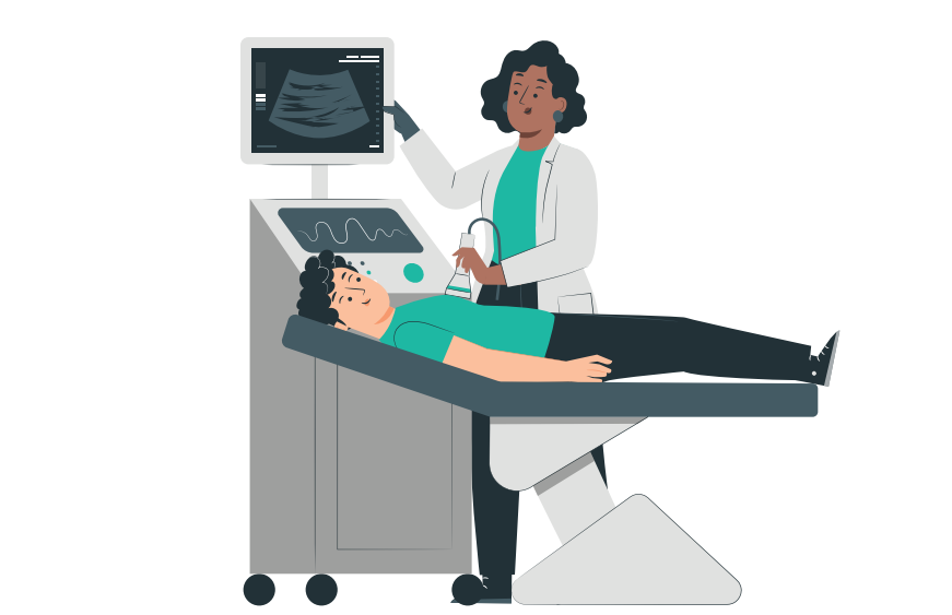
- The cardiology technician will assist you onto the Echo couch, where you will lay flat. It will be important to lie still and not talk during the procedure, to not interfere with the imaging. The cardiology technician will put gel on the ultrasound wand (transducer), to provide better images. The wand will be pressed firmly against the skin and moved over the chest area. The wand sends sound waves through the chest to the heart, recording the sound waves that bounce back from the heart. A computer changes the information into moving images, which are seen on the monitor. During the test, you may be asked to breathe in a certain way or to roll onto your left side.
- Fetal Echocardiogram is done similarly to the standard Transthoracic Echocardiogram, except the wand moves over the pregnant woman’s belly.
- A Stress Echocardiogram is completed in the same way as a standard Transthoracic Echocardiogram, except the images are taken before and after exercise. A Stress Echocardiogram often involves walking on a treadmill while an ECG is done.
- Your Echocardiogram should take between 15 and 60 minutes. Once the imaging has been completed, the cardiology team member will wipe off any gel that was used. They will assist you off the couch and you may get changed into your clothes.
- You will be escorted back to reception to exit the department/hospital. Your Echocardiogram report will be sent to the ordering physician within 48 hours. Please ensure you have your follow-up appointment booked after.
The Procedure for a Transesophageal Echocardiogram
This is what you can expect when you come to Health City for a Transesophageal Echocardiogram.
STEP 1 When you arrive at Health City, you will check in at reception.
STEP 2 A healthcare provider will lead you to the Echo room where you can change into a hospital gown. A nurse will check your vitals such as blood pressure, pulse, and oxygen levels.
STEP 3 The cardiology technician will assist you onto the Echo couch, where you will lie flat on your back. The Cardiologist will guide a thin, flexible tube called a TEE probe into your mouth, down your throat, and into your esophagus. The TEE probe goes through the esophagus and moves near the heart, sending sound waves through the chest to the heart, and recording the sound waves that bounce back from the heart. A computer changes the information into moving images, which are seen on the monitor.
STEP 4 Most Echocardiograms take less than an hour. After the Echo is completed, you may change into your clothes. The cardiologist will come to speak with you and will ensure you have a follow-up appointment in the coming days to review all images. Once all your paperwork is completed, you will be discharged and may leave the hospital.
Frequently Asked Questions
Other Patient Pathway Posts
Pulmonary Embolism

What to Expect After a Head Injury

Recovering After A Concussion
How to Care for a Child Who Has Croup
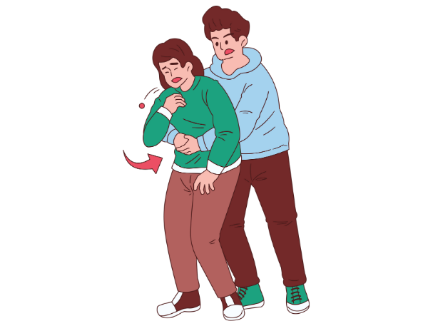
Choking: What You Can Do to Help
Understanding Your Headache & When to Seek Help
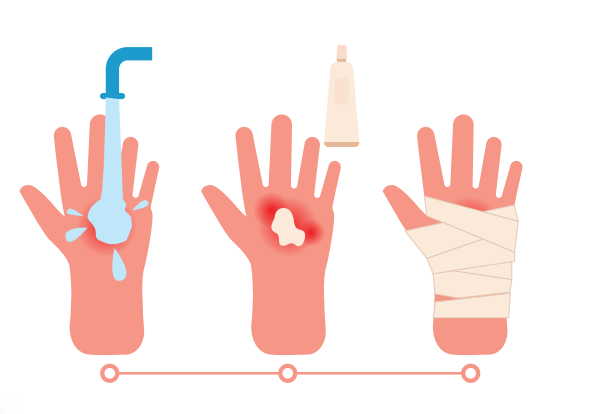
Understanding Burns
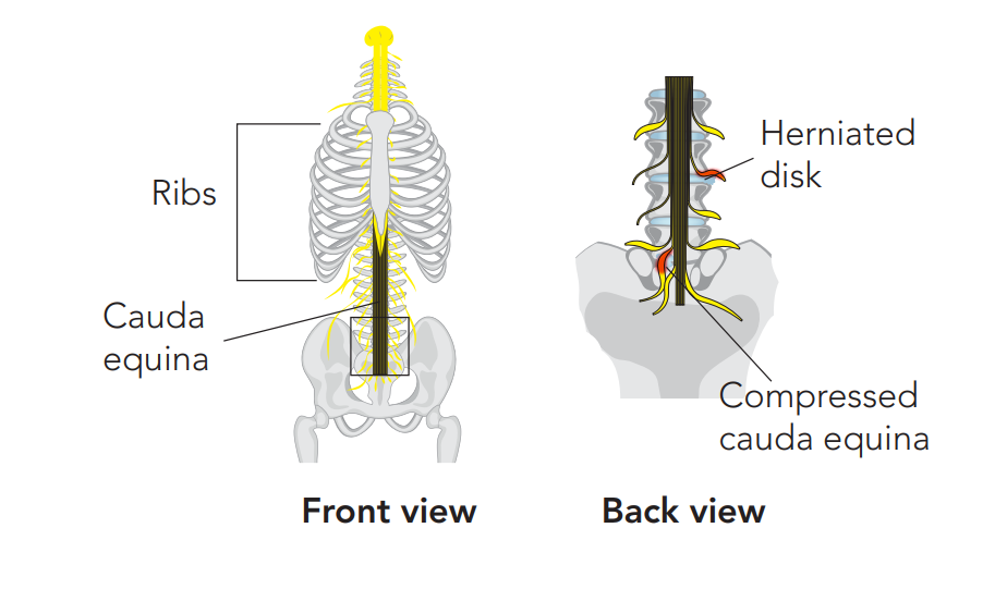
Recognising the Red Flags of Cauda Equina Syndrome
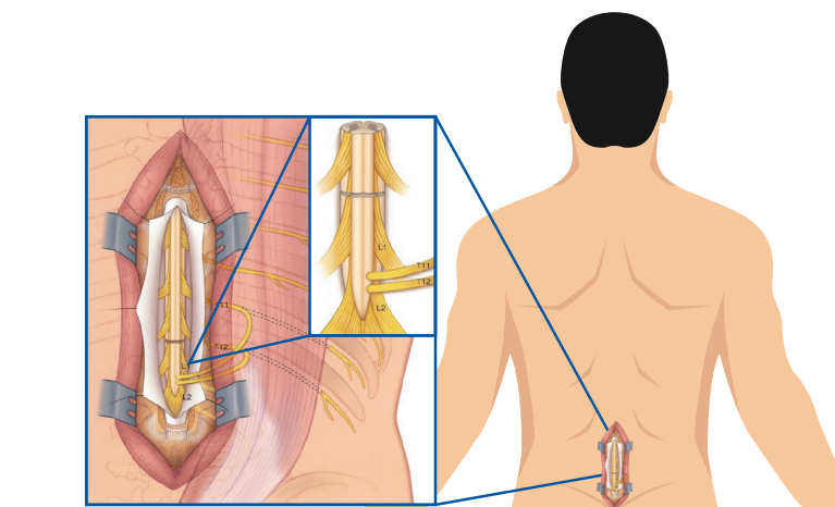
Recovering After Cauda Equina Syndrome
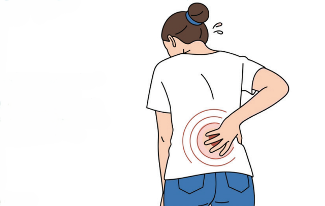
Recovery From Back Pain


